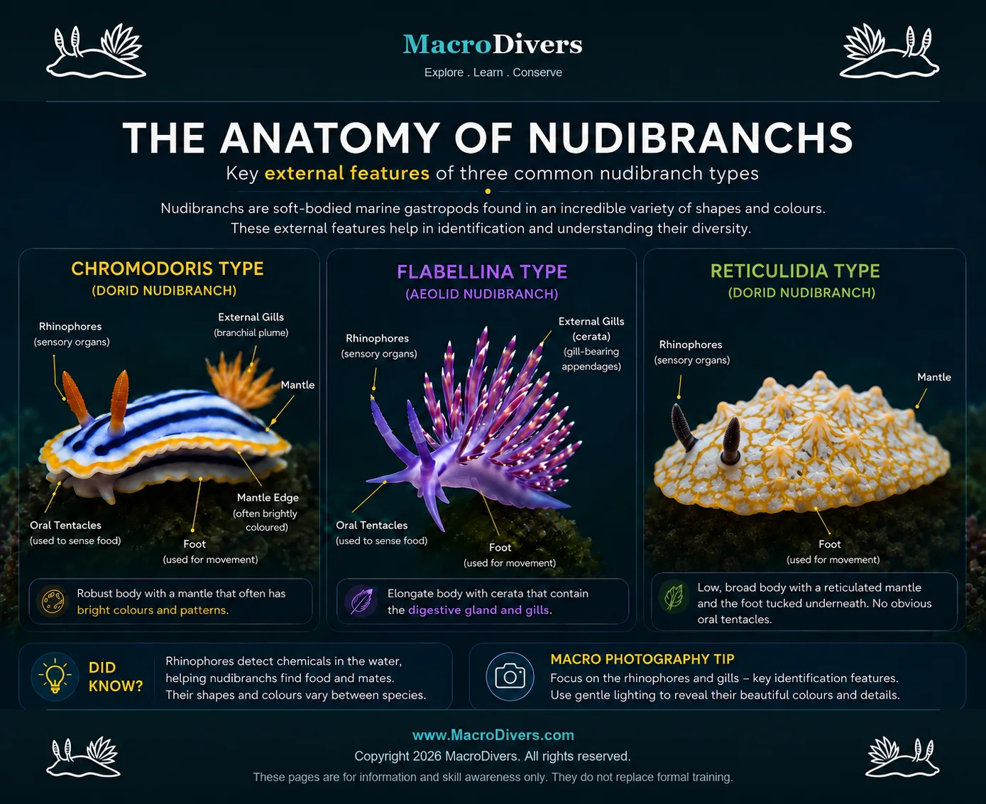
Nudibranchs may be small, but their bodies are full of useful clues.
Learning a few key features makes it much easier to understand what
you are seeing underwater and to identify species later from
photographs.
Rhinophores
What they do: Rhinophores are paired sensory
organs on the head. They help detect chemicals in the water,
including food, mates and surroundings.
Photo tip: If the rhinophores are sharp, the
image often feels much stronger.
Compare ringed, smooth, rolled and branched forms in the
MacroDivers rhinophore guide.
Rhinophore pockets
Many dorid nudibranchs can retract their rhinophores into
protective pockets when disturbed. This helps protect delicate
sensory structures.
Oral tentacles
Oral tentacles sit around the mouth area and help the animal
explore food and nearby surfaces. They can be important
identification clues in some groups.
Mouth and radula
The mouth is on the underside near the front. Nudibranchs feed
using a specialised rasping structure called a radula, rather
like a tiny toothed ribbon.
Foot
The foot is the muscular underside used for crawling. It
secretes mucus, allowing the nudibranch to glide slowly across
reef, sand, rubble or algae.
Mantle
The mantle is the upper body surface. In many nudibranchs it
carries the colours, spots, patterns and edges used for
camouflage, warning and identification.
Mantle edge
The outer edge of the mantle may be smooth, frilled, folded or
brightly coloured. It is often one of the first pattern areas to
compare when identifying species.
Digestive gland
The digestive gland helps process food and nutrients. In some
nudibranchs, colour and internal structures can be linked to
what the animal eats.
Gills
Gills are the respiratory structures sea slugs use to extract
oxygen from seawater. In many nudibranchs they appear as a
feather-like plume on the back, often surrounding the anus. In
other groups, respiration may be through cerata, hidden gills
beneath the mantle, or gas exchange across the body wall.
Want to learn more? See our
guide to nudibranch gill types.
Gill pocket
Some nudibranchs can retract their gills into a protective
pocket. This is useful when threatened or when moving through
tight spaces.
Anus
In many dorids the anus is near the rear of the body, close to
or within the branchial plume area. Its position helps explain
the arrangement of the gills.
Reproductive opening
Nudibranchs are hermaphrodites, meaning each individual has both
male and female reproductive organs. The reproductive opening is
usually on the right side of the body.
Why anatomy matters for identification
For divers and photographers, anatomy is not about memorising
Latin terms. It helps you know what to photograph. Clear images of
the rhinophores, gill plume, mantle edge, colour pattern and body
shape can make later identification much more reliable.